What is porphyrin in rats?
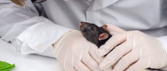
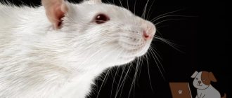
Have you noticed any unusual discharge from your pet's nose or eyes? This is a red-brown liquid with a consistency similar to blood. You may think that your pet has suffered an injury, such as a cut. If you want to treat the wound and find out the cause of the discharge, you will see that there is no wound. Such secretions in rats are called porphyrin.
The Veterinary Encyclopedia has a definition of this disease. Porphyrin - in a nutshell - is a red or burgundy-colored substance that resembles blood in appearance and composition. When porphyrin is released, you might think that your pet is crying tears of blood or has bloody snot.
Porphyrin forms on the animal's face near the nose or eyes. These bloody secretions are produced by special glands (Garderian), which are located in the corners of the eyes from the inside.
What does porphyrin look like?
By examining this substance under a microscope, it can be determined that it is similar to blood. It is an organic compound with four pyrol rings. Thus, porphyrin is a group of organic substances that have been identified in the wild. A property of porphyrins is the ability to bind metals.
How to solve this problem?
An increase or sudden release of red tears is a symptom of a specific problem. If your rat is not healthy, you should take her or him to a veterinarian for a health check.
During the physical exam, your veterinarian will check the front teeth, listen to the heart, lungs, and examine any other problems. Regardless of whether concerns arise during the health check, you should follow these recommendations:
- Reduce stress in the home and try to keep noise levels low.
- Make sure your rat's housing is the right size.
- Provide your pet's cage with toys and various entertainment items that will allow your pet to relieve stress.
- Check the filler material, avoid dusty fillers.
- Do not smoke near pets.
- Make sure there is good quality animal food and enough for everyone to avoid competition.
- Provide fresh water and change it daily.
Is it dangerous?
You can find out whether it is blood or porphyrin yourself. Take some of the substance onto a cotton pad and apply a drop of hydrogen peroxide on it. If the substance begins to hiss and changes color, it means there is blood on the cotton wool.
During a stressful situation or when the rat experiences pain, porphyrin is released in increased quantities. Up to one year, the animal produces a larger amount of this substance than an older rat.
By the age of two, very little fluid is produced by the glands. Often, such discharge is just a natural process, and the intervention of specialists is not required.
But there are times when the help of a veterinarian is necessary:
- liquid is constantly released from the rodent’s nose and eyes in large quantities;
- there is a suspicion that the rodent has mycoplasmosis, diphtheria or salmonellosis;
- if your pet has become slower, does not eat or drink well, or lies down in strange positions.
Symptoms of respiratory diseases
A bloody nose in a pet rat is one of the signs of a respiratory disease. Let us highlight its main symptoms:
- cough;
- wheezing;
- discharge of mucus from the mouth and nose;
- increased release of porphyrin;
- wheezing and gurgling when breathing;
- lethargy;
- lack of appetite;
- dyspnea;
- labored breathing.
Effect of porphyrin
Porphyrin lubricates the nictitating membrane and protects the animal's eyes from bright light. To put it simply, thanks to porphyrin, the animal can blink. Studies have found that in bright light conditions this substance is produced in larger quantities in rats.
The discharge may also increase in response to irritants such as blue light and ultraviolet light.
Also, the volume of secretions increases as the rat grows - the older it is, the more porphyrin. An individual one year old has more such secretions than, for example, a younger animal. And also, on the contrary, with age closer to two years, the production of this substance begins to decline again, and it is already formed in less than a year.
Prevention
When a rat has no problems and is not sick, its eyes, nose and mucous membranes are absolutely clean. When there is porphyrin in your pet's nose (for no reason), your pet requires the attention of a specialist. At the veterinary clinic, they will take tests and examine the animal, as a result of which it will be possible to understand whether everything is normal or whether there is a disease. In the latter case, the doctor will prescribe treatment at home or prescribe procedures at a veterinary institution.
To prevent the animal from developing bad symptoms, the owner must follow the following rules:
- the cage with the pet should be located in a quiet, calm place - the animal needs comfort;
- The animal should not be stressed, make noise, shout or wake it up abruptly - this will scare it;
- the rat must be fed healthy and wholesome food;
- You constantly need to monitor your pet, its condition, skin and mucous membranes.
Treating rats at home
First you need to determine whether your pet is healthy or sick. You can check this yourself, at home. We take cotton wool, collect the discharge from the animal’s face, and then drop a few drops of hydrogen peroxide on it. Let's see if the liquid has changed color. If, under the influence of hydrogen peroxide, the substance begins to hiss and change color, then it is blood, which means that the animal’s skin or mucous membranes have been damaged as a result of injury. If no reaction occurs and the color does not change, then it is porphyrin.
There are also ways to check discharge, for example, using ultraviolet light. You need to put a cotton pad with the substance under an ultraviolet lamp. If the drop starts to glow pink, then it is porphyrin.
Discharge from the ear, or otorrhea
Allergy
Fungus
15397 June 24
IMPORTANT!
The information in this section cannot be used for self-diagnosis and self-treatment.
In case of pain or other exacerbation of the disease, diagnostic tests should be prescribed only by the attending physician. To make a diagnosis and properly prescribe treatment, you should contact your doctor. Discharge from the ear: causes of occurrence, what diseases cause it, diagnosis and treatment methods.
Definition
Earwax is a physiological secretion from the ear canal and protects the hearing aid from pathogenic bacteria. It contains lard, fatty acids and fat-like substances, as well as various mineral salts. Normally, a person produces 15–20 mg of earwax per month, which has the appearance of a sticky yellow-brown mass. All other discharges are considered pathological and indicate ear diseases.
Types of ear discharge
The discharge may be clear, white, light or dark yellow, or greenish (if there is pus). If blood enters the ear secretion, the discharge becomes reddish or brownish in color.
The consistency of the discharge may be watery, have a cheesy or flaky texture, and sometimes crusting may form.
An unpleasant smell of discharge due to the presence of pus in it can serve as a diagnostic sign.
What diseases and conditions cause discharge from the ear ? Wax plugs
. Excessive work of the sulfur glands leads to the formation of sulfur plug. Most often, this problem occurs in patients with diabetes mellitus, metabolic syndrome, and high cholesterol levels in the blood. The formation of cerumen plugs is provoked by increased viscosity of sulfur, dry skin, the entry of small foreign particles into the ear (for example, industrial dust), as well as excessive hair growth in the ear canal. Often, wax plugs are observed in people involved in water sports, using hearing aids, and miniature headphones.
With improper hygiene measures and independent attempts to remove excess earwax, there is a risk of pushing it deeper into the ear canal, thereby causing the formation of a plug.
Clinical signs of cerumen plug are pain and congestion in the ear, tinnitus, especially painful when the cerumen comes into contact with the eardrum, sometimes headache, dizziness, and nausea.
Mucopurulent and purulent discharge is a symptom of inflammation of the outer and middle ear. For inflammation of the outer ear (otitis externa)
the pathological process can develop in the auricle and external auditory canal (up to the eardrum). Most often, otitis externa occurs due to infection of the ear by bacteria and microscopic fungi. Its first signs are, as a rule, pain in the ear, itching, and, less often, hearing loss and a feeling of fullness. Mucopurulent discharge appears only with a widespread form of the inflammatory process throughout the entire ear canal.
The source of purulent discharge in the outer ear can also be a boil
located in the concha or ear canal.
With otitis media of the middle ear
, mucopurulent and purulent discharge becomes the result of infection of the sterile effusion from the inflamed tissues of the ear. Since the chamber of the middle ear is closed by the eardrum, purulent discharge can appear in the outer ear only after a hole has formed in it. This is preceded by severe pain in the ear, fever, decreased hearing, and in children - overexcitement, sometimes vomiting.
For mastoiditis
(inflammatory lesion of the mastoid process of the temporal bone), purulent discharge from the ear also appears. As a rule, this disease develops as a complication of otitis media and is accompanied by fever, pain and swelling in the mastoid area behind the ear.
Transparent bloody or purulent discharge appears with acute infectious myringitis
(inflammation of the eardrum), which may be of fungal or bacterial origin. Blisters filled with blood form on the surface of the eardrum, which then burst. In addition to the discharge, ear congestion is observed.
Transparent, colorless or slightly pinkish discharge from the ear can be a consequence of liquorrhea - the leakage of cerebrospinal fluid. It enters the auricle during fractures of the skull bones
(usually temporal) due to injury.
In addition, clear watery discharge sometimes accompanies allergic otitis
, which is also characterized by other symptoms - itching, ear congestion.
Unchanged blood appears from the ear, usually after injury and rupture of the eardrum
.
Such injury can occur after acoustic and mechanical shocks, as well as as a result of improper hygiene procedures. A ruptured eardrum is always accompanied by severe pain.
The appearance of bloody-purulent discharge from the ear is one of the indications of the presence of a polyp on the eardrum or the mucous membrane of the middle ear
. A polyp is a growth of tissue in response to irritation. The appearance of a polyp is preceded by active inflammation of the middle ear. In addition, polyps can be a consequence of myringitis, otitis externa, or malignant neoplasms. By perforating the eardrum, the polyp can protrude into the external auditory canal, leading to hearing loss.
Minor discharge, sometimes forming crusts and having an unpleasant odor, is characteristic of cholesteatoma.
– a tumor-like formation formed from the epidermis of the ear canal.
In most cases, cholesteatoma complicates chronic purulent epitympanitis
and is formed from layers of keratinized epidermis, water, proteins, fats and cholesterol. The formation of cholesteatoma is accompanied by feelings of heaviness and fullness in the ear, and headache. If left untreated, it can gradually invade the mastoid process and cranial cavity.
For otomycosis
loose cheesy discharge is observed. The main culprits of the disease are molds (usually localized in the outer ear) and yeast-like fungi (usually found in the middle ear). Clinical signs of otitis externa in these cases include pain and a colored, cheesy-necrotic discharge from the ear. Patients complain of tinnitus and dizziness.
A discharge that contains large, greasy flakes, sometimes mixed with pus, is characteristic of seborrheic ear dermatitis.
. The disease can affect not only the ear, but also the scalp. Clinical signs include severe itching, swelling of the ear, peeling skin and weeping wounds.
Transparent discharge mixed with blood (bloody discharge) indicates bullous, or influenza, otitis media.
. Bullae (bubbles of fluid) appear on the surface of the ear canal and eardrum. When they burst, the liquid with ichor flows through the ear canal into the auricle.
Which doctors should I contact for ear discharge?
In most cases, ear diseases are characterized by a clear clinical picture, which is based on pain. Such patients are treated by an otolaryngologist.
In the presence of a traumatic brain injury, which is accompanied by liquorrhea, urgent hospitalization is necessary. Otherwise, an unfavorable prognosis is possible.
Discharge from the ear is not always accompanied by pain, in particular with allergic otitis media, which is treated by, and.
Diagnosis and examinations for ear discharge
If the formation of a cerumen plug is suspected, the doctor performs an otoscopy, during which an accumulation of sulfur in the ear canal is detected. Diagnosis of external and otitis media is carried out on the basis of patient complaints, otoscopy, and palpation of the parotid area. It is recommended to culture the discharge from the ear to determine the causative agent of the disease and its sensitivity to antibiotics. Audiometric examination is possible.