If your guinea pig's eyes suddenly start to water, this can have various causes, from injury to disease. Eye diseases in guinea pigs are quite common. There are many reasons for their development in this animal species:
- bacterial infections;
- injuries (injections with hay, scratches received during the division of territory);
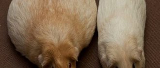
- metabolic disorders (obesity, excess or deficiency of vitamins);
- as a secondary disease, for example, against the background of diabetes mellitus;
- biological old age of the animal.
Main causes of eye diseases
Ophthalmic diseases often plague guinea pigs. Although a rodent can lead a full life even if it is blind, you should not ignore situations when its eyes begin to water and turn red. These are the first symptoms that should alert the animal owner. Early treatment will help preserve vision.
The main reasons why guinea pigs have watery or purulent eyes:
- allergy to the filler;
- lack of vitamins A and C;
- infectious eye lesions;
- autoimmune diseases;
- eye injuries;
- disorders of the endocrine system, diabetes mellitus;
- pathological structure of the eyelids (congenital anomalies);
- entry of a foreign body into the eye - debris, dust.
Symptoms of pharyngitis
The first signs of the disease may differ depending on the type of pharyngitis. They are both local and general in nature. But there are common signs that are characteristic of any type of pharyngitis: sore throat, bad breath, stuffy ears and difficulty swallowing. With an active inflammatory process, an increase in body temperature above 38° can be observed - this is how the body fights a foreign infection. General signs: sweating, poor appetite, weakness, dizziness, fatigue, fever, chills. Some complain of pain and noise in the ears, and discomfort when exposed to loud sounds.
- During acute catarrhal pharyngitis, swelling and redness of the mucous membranes of the larynx appears. Also, red follicles may form on the back wall of the throat, and clear and slightly cloudy mucus may accumulate. There is swelling and redness of the tongue.
- In the purulent form of acute pharyngitis, ulcers with an accumulation of purulent masses appear on the surface of the posterior pharyngeal wall.
Eye diseases in guinea pigs
Not all, but most eye diseases in guinea pigs are accompanied by tearing and redness of the eyelids. Such symptoms indicate inflammation.
Conjunctivitis
Conjunctivitis in guinea pigs is the most common ophthalmological disease. The inflammatory process affects only the mucous membrane of the eyeball - the conjunctiva. The disease can affect one or both organs of vision.
Inflammation develops for several reasons:
- Due to pathogenic microorganisms that have entered the eye - fungi, bacteria or viruses.
- Against the background of allergies. Guinea pigs often have a reaction to the components of the litter, dust, tobacco smoke or cleaning and detergents.
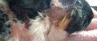
The very first sign of conjunctivitis is that your pig's eyes are watery. Further, slight redness and swelling of the eyelids is observed. The rodent squints and rubs its muzzle with its paw, which only aggravates the problem. Then pus will appear. The discharge will become thicker and turn yellow. The eyelids will stick together. At this stage, the eyes are no longer watery, but the rodent experiences severe discomfort.
It is important to start treatment as quickly as possible. Ideally, the owner should react when the eyes just begin to water. Advanced conjunctivitis can lead to complications - inflammation of the cornea and even loss of vision.
Attention! Sometimes inflammation of the conjunctiva occurs against the background of deadly infectious diseases. The eyes of guinea pigs become watery and fester due to pasteurellosis. If, in addition to conjunctivitis, there is a runny nose, loss of appetite, depression and diarrhea, you should immediately put the animal in a separate cage and show it to a veterinarian.
Keratitis
This is an inflammation of the cornea (sclera) of the eye. Reasons for the development of keratitis:
- complication after untreated conjunctivitis;
- injury;
- entry of a foreign body;
- allergy.
Keratitis causes severe discomfort to the guinea pig. The affected eye is watery and painful. Because of this, the rodent constantly squints and rubs its muzzle with its paw. In bright light, the discomfort intensifies. The guinea pig becomes restless and loses its appetite.
If the inflammatory process affects the deeper layers of the cornea, scars will remain on it. This will lead to deterioration of vision or the appearance of a cataract.
Cataract
This is one of the most common causes of complete or partial loss of vision. In most cases, cataracts in guinea pigs develop at an advanced age. As the animal ages, the lens loses its properties and becomes cloudy. It can no longer perform its function of transmitting and refracting light.
Reference. With cataracts, the animal sees the image blurred, as if looking through a stream of water.
If cataracts are diagnosed in a young rodent, then its development is associated with other reasons:
- heredity;
- infectious diseases;
- eye injury;
- metabolic disorders.
With cataracts, the eyes do not water, the mumps does not have purulent discharge or discomfort.
At the early stage of the disease, the animal behaves as usual. An attentive owner may notice a slight change in the color of the lens around the perimeter. In this case, the rodent can freely navigate in space.
The disease gradually progresses, then cloudy spots appear in the central part of the lens. Now the animal sees only the outlines of objects. At the next stage, the entire biological lens becomes cloudy. Now the guinea pig is almost blind. She can only see light. At the final stage, the fibers of the lens begin to liquefy, after which it acquires a milky color.
Cataracts cannot be cured with medications. The only way to restore vision is surgery. However, such an expensive operation is almost never performed on guinea pigs.
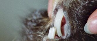
Belmo
A cataract is a cloudy spot on the cornea of the eye, which usually appears at the site of a scar. In guinea pigs, it can form after an eye injury or any inflammatory processes that affect the cornea.
A guinea pig cataract looks almost exactly like a cataract. A cloudy porcelain-white spot appears on the eyeball. At the same time, the animal’s vision decreases.
Unlike cataracts, a cataract is accompanied by a burning sensation. The eyes water and the inside of the eyelid turns red.
If left untreated, the thorn will grow in size over time. This will lead to complete or partial loss of vision.
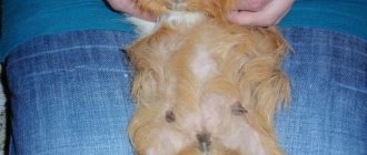
Fat eye
This is what people call prolapse of the conjunctival sac. This is a fatty fold under the eyelid that seems to run over the eyeball and can close part of the pupil. An oily eye does not water or hurt. This pathology does not pose any danger to the health of the guinea pig. However, fatty tissue may obscure the view.
In this case, the protruding fold is removed with a laser.
Important! Protrusion of fatty tissue from under the eyelid is inherited. Therefore, individuals with such a defect are not allowed for breeding. It is believed that cream and black guinea pigs are more susceptible to this pathology.
Glaucoma
This disease progresses rapidly and leads to complete blindness. Glaucoma is characterized by destruction of the retina as a result of increased eye pressure. The optic nerve gradually atrophies, so it cannot transmit signals to the brain. Glaucoma is more common in older guinea pigs.
Symptoms:
- swelling and redness of the cornea;
- watery eye;
- photophobia.
Glaucoma causes pain in the guinea pig's eye. The rodent develops anxiety, which intensifies in bright light. At the initial stage of the disease, you can get by with the use of drops that stabilize eye pressure. In advanced cases, you will have to resort to removing the organ of vision.
Retrobulbar abscess
With this disease, a cavity with pus forms in the retrobulbar space. An orbital abscess in a guinea pig can occur due to dental problems.
The clinical picture is as follows:
- swelling and hyperemia of the eyelids;
- exophthalmos;
- the eyeball partially loses mobility and protrudes greatly;
- the rodent looks lethargic;
- body temperature rises;
- When you open your mouth, the pain intensifies, so the animal squeaks.
Attention! The abscess can spontaneously open, then its contents will come out through the conjunctiva. However, there is a high probability of the abscess breaking inside. In this case, the development of phlegmon of the orbit of the eye, infection of the meninges and sepsis is possible.
Retrobulbar abscess is treated surgically. The doctor can remove the pus with a needle, rinse the abscess cavity with a disinfectant solution, and prescribe broad-spectrum antibiotics. In advanced cases, the organ of vision has to be removed.
Entropion
With entropion, the eyelid is turned towards the eyeball. This disease is often found in Rex, Texel and Teddy guinea pigs. During movement, the eyelid constantly contacts the cornea and irritates it, so the eye waters. At the site of microdamage, ulcers and then cataracts may subsequently form.
Entropion often disappears as the pet gets older. If this does not happen, a simple operation is performed to eliminate the defect.
White discharge from the eyes
Guinea pigs normally secrete a small amount of white fluid from their eyes, which resembles milk. This is the secretion of the Harderian glands. It is designed to prevent the eyeballs from drying out. There is no reason to worry if there is little discharge, and the rodent has a good appetite and looks healthy.
When the secretion increases and the animal’s general health worsens, you need to contact a veterinarian.
Forms and types of cataracts
According to the nature of origin, congenital and acquired forms of leukoma are distinguished. Congenital occurs as a result of transplacental infection. Transmitted from mother to fetus. In the acquired form, the formation of a cataract occurs due to external factors.
According to the severity of turbidity, they are distinguished:
- cloud - minor changes;
- corneal spot - partial spread of leukoma;
- thorn - dense and extensive localization.
The cataract can be flattened, convex, or fused to the iris.
According to its location, the leukoma can be central, when the cataract completely or largely covers the pupil. Total - the cornea is completely closed. In these cases, visual acuity is significantly reduced. With peripheral leukoma, the pupil is not affected by clouding and vision does not decrease.
Treatment of tearing in guinea pigs
If you notice any strange symptoms in your pet, be it redness of the eyelids, swelling or purulent discharge, it is recommended to seek veterinary help. Treatment of most ophthalmological diseases is possible only under the supervision of a doctor.
The most common reason why guinea pigs' eyes become watery and purulent is conjunctivitis. Drops and ointments are used to treat it.
The appearance of purulent discharge from the eyes is a clear sign of a bacterial infection. In such cases, drops are prescribed:
- Levomycetinaceous;
- Tsiprovet;
- Tobrex.
Also, for tearing and purulent eyes, you can use Tetracycline ointment. It is applied under the lower eyelid in the morning and evening for 5-7 days.
Before instilling the medicinal composition, it is recommended that the pig's eyes be washed. To do this, you will need several cotton pads and saline solution. If you don’t have it on hand, you can take warm boiled water. The sponge is moistened, lightly squeezed and wiped first one eye.
It is important to remove all crusts on the eyelids and clean the fur around them from pus. Next, take a clean cotton pad and do the same on the other side.
For corneal injuries, the drug Balarpan is used. It is used a quarter of an hour after drops with an antibacterial effect. Then, after another 20 minutes, the regenerating gel Solcoseryl is applied.
First aid
call an ambulance immediately . Doctors may decide on emergency hospitalization if the patient has difficulty breathing, a swollen tongue, or symptoms indicating intestinal damage.
What to do before the doctors arrive?
First aid, which should be provided at the first symptoms of angioedema in children, involves clearing the airways, checking breathing intensity, heart rate, and blood pressure. Sometimes it is necessary to perform cardiopulmonary resuscitation, so after the first case, parents are recommended to take first aid courses. At the final stage, medications should be administered to the child.
First aid equipment includes:
- glucocorticosteroids (“Prednisolone”, “Dexamethasone”);
- antihistamines;
- adrenalin.
Sequence of administration: adrenaline, then glucocorticosteroid, then antihistamine. If the reaction is moderate, then adrenaline is excluded.
| Adrenalin | It is injected intramuscularly into the thigh (middle third outside) at the rate of 0.01 mg for each kg of the child’s weight. If there is no effect, then the injections are repeated every 15 minutes. |
| Glucocorticosteroids | Injected intramuscularly into the buttock or intravenously. You can pour the medicine from the ampoule under the tongue - this way the effect will come faster. The dosage of Prednisolone is from 60 to 150 mg, Dexamethasone is from 8 to 32 mg. |
| Antihistamine | Intramuscular injection is preferable. It is possible to take a pill, but the effect will come later. The dosage depends on the medicine. For example, “Loratadine” – 10 mg, “Cetrizine” – 20 mg. |
Important! In case of swelling of the larynx, a tracheostomy is performed urgently.
Diagnosis of pharyngitis
Detection of all types of pharyngitis begins with a visual examination of the larynx using a special device and taking an anamnesis. A throat swab is also taken for examination to test for diphtheria.
Other types of diagnostics:
- Cultural examination - inoculation of taken materials on a nutrient medium.
- Rapid diagnosis - identification of streptococcal antigen in throat swabs.
- Immunoserological diagnosis - the method is used in case of streptococcal infection.
Laboratory research:
- Complete blood count – exclusion of blood diseases, infectious mononucleosis;
- A general urine test helps rule out kidney disease (glomerulonephritis).
Depending on the symptoms of the disease, as well as the condition of the larynx, the presence or absence of cough, fever, plaque on the tonsils and soreness and increased size of the lymph nodes, additional consultations with other specialists may be necessary: an endocrinologist, a cardiologist, an allergist.
How does pharyngitis manifest and progress in children?
Children suffer from pharyngitis more severely than adults. This especially applies to babies under one year old. Swelling of the mucous membrane can cause signs of suffocation; the pain that accompanies the disease reduces the child’s appetite. Often, a baby’s body temperature can reach 40°. The most difficult thing in this situation is that a small child cannot say what hurts.
Incorrect treatment can lead to irreparable consequences for a small, fragile organism. Therefore, at the first signs of pharyngitis, consult a doctor immediately.